


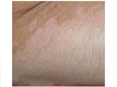
Hypopigmentation on arm from vitiligo
Vitiligo is caused by the loss of pigment-producing cells in the skin (melanocytes), which, in turn, reduces the production of melanin not only in skin but also in mucous membranes, eyes, hair bulbs, and in the ears, leading to alterations in both structure and function of these organs.
It is seen clinically as depigmented, welldemarcated macules with variable progression ranging in size from 5 to 50 mm. Lesions may be found anywhere on the body. However, initial lesions are most frequently found on the hands, forearms, feet, face, and lips. In some cases, these patches can appear all over the body. The white patches tend to be sharply circumscribed and are quite striking in darker skin, which can be cosmetically very disabling. There may be increased pigmentation of surrounding skin. They may at first be few in number but tend in somewhat erratic fashion to increase in time.
Family history of vitiligo is established in 25 to 30 % of patients. Affected families have an increased incidence of graying of the hair.
Onset is often insidious, but is frequently related to a recent stress, illness, or trauma (e.g., sunburn). It can occur at any age, but in nearly 50% of patients peak onset is in the second and third decades of life. It is relatively common affecting about 1-2% of the population in all races, ethnicities and genders.
Childhood vitiligo is seen in children under 12 years and is different from the other type in that depigmentation frequently occurs in long segments.
Sometimes spontaneous remission occurs and repigmentation is seen.
It is thought to be a result of immunologic, genetic, and neurogenic reasons but the precise pathogenesis remains conjectural. Vitiligo may be associated with other autoimmune diseases, especially thyroid disease and diabetes mellitus. Other associated autoimmune diseases include pernicious anaemia, Addison disease, and alopecia areata. Localized vitiligo is restricted to one area including a segmental or quasi-dermatomal distribution.
Diagnosis of vitiligo is based almost exclusively on the clinical examination.
There are four main types of vitiligo: generalized, acrofacial, segmental, and universal.
Generalized is the most common type. Itinvolves greater than 10% of the body surface area. Patients present with bilaterally symmetric lesions involving the peri-orofacial areas, neck, torso, bony prominences of hands, wrists, and legs, extensor surfaces, orifices, axillae, and mucosal areas.
Acrofacial vitiligo presents with lesions on distal fingers and on the facial orifices.
Segmental vitiligo presents in an asymmetric dermatomal distribution. It has an earlier age of onset and is not associated with autoimmune diseases. It has a poorer prognosis for treatment.
Universal vitiligo, which has been associated with multiple endocrinopathies, presents with depigmented macules and patches involving almost the entire body.
There is no cure for vitiligo. Treatment may include covering smaller patches with long-lasting dyes, light-sensitive drugs, in addition to ultraviolet A light therapy, corticosteroid creams, and depigmentation of the remaining skin. The greatest way to lower your chances of developing these skin pigmentation disorders is to be careful when in the sun, and remember to wear a good sunscreen , light clothing, and stay out of the sun when the rays are the most intense.
>
>
>
>
>
>
>
>
>
>
>
>
>
>
>
>
>
>
>
>
>
>
>
>
>
>
INTRODUCTION
PIGMENTARY DISORDER TYPES
Disorders of
Hyperpigmentation
Disorders of Hypopigmentation
COMMON PIGMENTARY DISORDERS
Age Spots/Liver Spots/Lentigos
Acanthosis Nigricans
Albinism
Cafe-au-Lait Macules
Ephelides (Freckles)
Erythema Dyschromicum Perstans (Ashy Dermatosis)
Familial Racial Periorbital Hyperpigmentation
Idiopathic Guttate Hypomelanosis
Leopard Syndrome
Linea Nigra
Melanoma
Melasma
Nevus (birthmarks/moles)
Parkinsons Disease
Phytophotodermatits
Pityriasis Alba
Poikiloderma of Civatte
Postinflammatory
Hyperpigmentation & Hypopigmantation
Seborrheic Keratoses
Sturge-Weber Syndrome
Substance Induced
Hypermelanosis
Tinea Nigra/Tinea Versicolor/Pityriasis
Versicolor
Vitiligo
Waardenburg Syndrome

<< Previous: Tinea Nigra / Tinea Versicolor / Pityriasis Versicolor
Next: Waardenburg Syndrome >>